0. Introduction
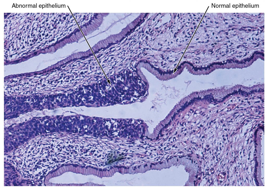
The cells found in the human body contain essentially the same internal structures yet they vary enormously in shape and function. The variation in cells is not randomly distributed throughout the body, rather, they occur in organized layers. Such aggregations of cells that are similar in structure and work together to perform a specialized function are referred to as tissues. The micrograph that opens this chapter shows the high degree of organization among different types of cells in the tissue of the cervix. You can also see how that organization breaks down when cancer takes over the regular mitotic functioning of a cell.
The human body starts as a single cell at fertilization. As this fertilized egg divides, it gives rise to trillions of cells, each built from the same blueprint, but organizing into tissues and becoming irreversibly committed to a developmental pathway.